
Glioma: what this diagnosis means and what the options are
"Glioma" is not one diagnosis. That single word covers tumours with radically different outlooks.
The most common brain tumour in adults — and the one whose outlook has changed most in the last decade.
A brain metastasis is a tumour that did not begin in the brain: it originated in another organ and reached the nervous system through the bloodstream. This differs from a glioma, which arises from brain tissue itself.
Metastases are by far the most common brain tumours in adults: they outnumber all primary brain tumours combined.
In a proportion of patients, the brain metastasis is the first manifestation of a cancer not yet diagnosed. In those cases, staging to locate the primary tumour forms part of the initial workup.
That last point deserves emphasis: not every brain lesion in an oncology patient is a metastasis. It may be an abscess, an independent primary tumour, a demyelinating lesion, or radionecrosis from previous treatment. Confirming the diagnosis changes treatment entirely.
Dexamethasone reduces oedema around the lesion and usually improves symptoms within hours or a few days. It is symptomatic rather than antitumour treatment, and is titrated to the minimum effective dose because of side effects.
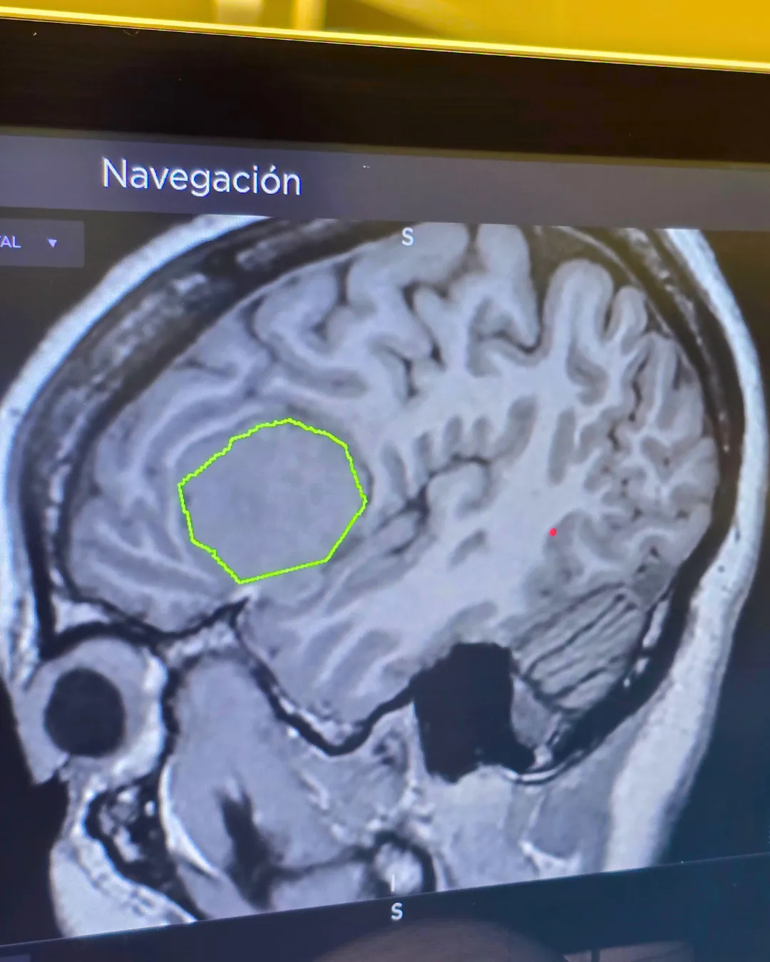
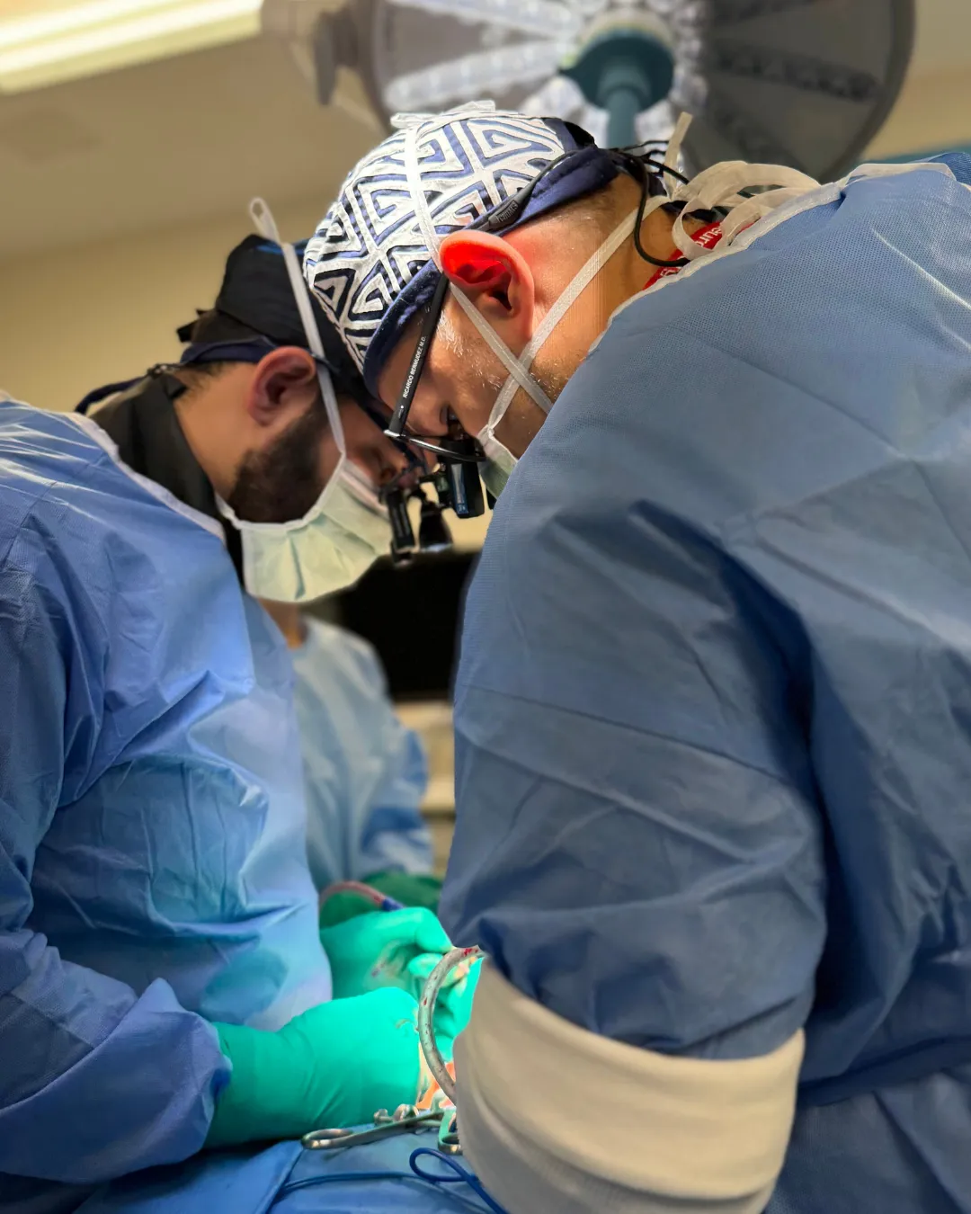
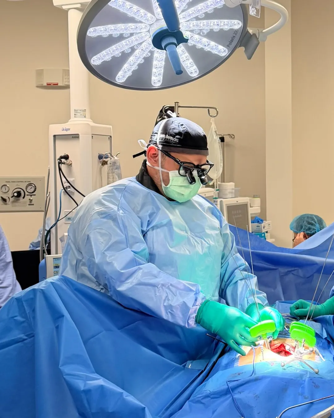
Resection is performed with neuronavigation, intraoperative neuromonitoring and magnification, to remove the lesion while preserving function. Metastases are usually better circumscribed than infiltrative gliomas, which often allows complete resection.
Delivers a high, highly precise dose of radiation to the lesion in one or few sessions, sparing surrounding healthy tissue. No incision or general anaesthesia is required.
It has become the reference treatment for small and multiple lesions, and is also used after surgery on the resection cavity to reduce local recurrence.
Irradiates the entire brain. Its use has reduced considerably because of effects on cognitive function, and it is now reserved for widely disseminated disease or specific situations. When used, hippocampal-sparing techniques reduce that impact.
Much of the last decade's change sits here. Several targeted therapies and immunotherapies can cross the blood-brain barrier and act on brain metastases, something classical chemotherapy achieved only with difficulty. The choice depends on tumour type and molecular profile.
Tumour-specific prognostic scales exist that oncologists use to guide decisions. What matters for patients and families is that estimates have improved significantly for several subtypes, and that figures found online usually come from series predating modern radiosurgery and current targeted therapies.
That conversation deserves to happen with the treating team, with the complete diagnosis available and enough time to ask everything that needs asking.
Yes, in selected cases. Surgery is considered when there is a single lesion or few accessible lesions, when they cause mass effect with significant symptoms, when histological diagnosis is needed, or when there is risk of herniation. It is weighed alongside the patient's functional status and control of systemic disease.
It is the delivery of a high, highly precise radiation dose to the lesion in one or few sessions, sparing healthy brain tissue. It requires no incision or general anaesthesia. It is today the reference treatment for small and multiple metastases, and is also used after surgery to reduce local recurrence.
There is no fixed number. Stereotactic radiosurgery can treat multiple lesions in a single session, and the limit is set more by total tumour volume, the size of each lesion and the patient's condition than by an exact count. The decision is made by a multidisciplinary board.
No. It may be an abscess, an independent primary tumour, a demyelinating lesion, or radionecrosis from previous treatment. When there is diagnostic doubt, a biopsy is performed, because treatment changes entirely depending on the real diagnosis.
No. Some are detected on surveillance imaging in asymptomatic patients, especially in cancer types where routine brain follow-up is performed. Small lesions away from functional areas may cause no clinical manifestation for some time.
Written and reviewed by Dr. Ricardo Bermúdez, neurosurgeon. Last reviewed: 2026-09-09.
General informational content. It is not medical advice, does not create a doctor-patient relationship, and must not be used to self-diagnose or to decide on treatment. Outcomes vary from person to person. If you have symptoms, see a doctor.
"Glioma" is not one diagnosis. That single word covers tumours with radically different outlooks.
In an older adult, a knock that seemed insignificant can cause symptoms six weeks later.
Most blows to the head have no consequences. The problem is knowing which ones do.