

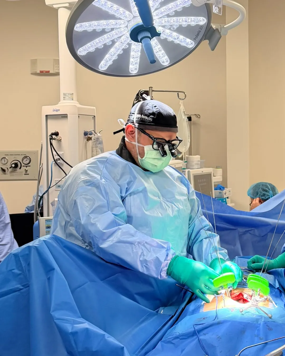
Traumatic brain injury: what to watch for in the first 48 hours
Most blows to the head have no consequences. The problem is knowing which ones do.
In an older adult, a knock that seemed insignificant can cause symptoms six weeks later.
A subdural hematoma is a collection of blood between the brain and the dura mater, the tough membrane covering it. Because it takes up space inside a skull that cannot expand, it compresses the brain.
What makes this condition particularly treacherous is the delay: in its chronic form, symptoms can appear weeks or even months after a knock that seemed unimportant at the time. Many patients do not even remember hitting their head.
| Acute | Subacute | Chronic | |
|---|---|---|---|
| Time since injury | < 3 days | 3 days – 3 weeks | > 3 weeks |
| Typical mechanism | Severe trauma | Moderate trauma | Minor or unremembered trauma |
| Patient profile | Any age | Variable | Older adults, anticoagulated |
| Course | Rapid deterioration | Progressive | Slow, insidious |
| Urgency | Immediate emergency | Urgent | Priority, not always immediate |
With age the brain reduces slightly in volume. That leaves more space between brain and skull, and the small bridging veins connecting them become stretched and exposed. A sudden head movement can tear one of those veins without any forceful impact.
Blood accumulates slowly. At first the available space absorbs it without symptoms. When the collection grows enough to displace the brain, symptoms begin.
Here the picture is far subtler and is frequently mistaken for dementia, depression or "just old age":
Non-contrast head CT is the investigation of choice. It is fast, available in the emergency department, and shows the collection clearly, including its approximate age from the density of the blood.
MRI is reserved for doubtful cases, isodense hematomas hard to see on CT, or bilateral collections that balance each other out and do not shift the midline.
A small hematoma without significant brain displacement, in a patient with no neurological symptoms, can be monitored with clinical and imaging follow-up. Many reabsorb on their own. If the patient is anticoagulated, the risk-benefit of that medication is reassessed together with their cardiologist or physician.
Burr hole drainage. The standard procedure for chronic hematoma. One or two small openings are made in the skull, the contents — usually liquid rather than clot by that stage — are drained, and the cavity is irrigated. It is a relatively short operation, well tolerated even in elderly patients.
Craniotomy. Needed for acute hematoma with solid clot, or when organised membranes prevent simple drainage. A larger bone window is opened to evacuate the clot and control bleeding.
Middle meningeal artery embolisation. A more recent endovascular technique that cuts the blood supply feeding the membranes of a chronic hematoma. It is used alongside surgery or as an alternative in selected cases, particularly to reduce recurrence.
Chronic subdural hematoma responds well to drainage: neurological improvement is usually rapid and many patients return to their previous state. The result depends above all on how long the brain was compressed before treatment and on the patient's general condition.
Recurrence is not rare — the subdural space remains dilated and can refill — and is more common in anticoagulated patients, marked brain atrophy, or organised membranes. Imaging follow-up after surgery is therefore part of the treatment, not an extra.
Acute hematoma carries a more guarded prognosis, closely tied to neurological status on arrival at hospital and how quickly it is evacuated.
The acute form presents within hours or days. The chronic form can take from three weeks to several months to cause symptoms, and often the patient does not even recall the injury because it may have been minor. That is why, faced with neurological changes in an older adult, it is worth asking about falls in recent months.
Yes. Small hematomas without brain displacement and without symptoms can reabsorb spontaneously with clinical and imaging monitoring. The decision to observe or drain depends on size, midline shift, symptoms and the patient's general condition.
Burr hole drainage is a relatively short, well-tolerated procedure often performed even in very elderly patients. The risk of leaving a symptomatic hematoma untreated is usually greater than the risk of surgery. Each case is assessed individually, considering comorbidities and anticoagulant medication.
This decision must be made jointly by the neurosurgeon and the doctor who prescribed the anticoagulant, usually a cardiologist or physician. It is generally paused around surgery and reintroduced afterwards according to individual thrombotic risk. It should never be stopped on your own initiative.
Worsening headache, vomiting, excessive drowsiness or difficulty waking, confusion, behavioural change, weakness on one side, difficulty speaking, visual disturbance, unsteadiness when walking, or seizures. Any of these signs means going to the emergency department.
Written and reviewed by Dr. Ricardo Bermúdez, neurosurgeon. Last reviewed: 2026-09-09.
General informational content. It is not medical advice, does not create a doctor-patient relationship, and must not be used to self-diagnose or to decide on treatment. Outcomes vary from person to person. If you have symptoms, see a doctor.
Most blows to the head have no consequences. The problem is knowing which ones do.
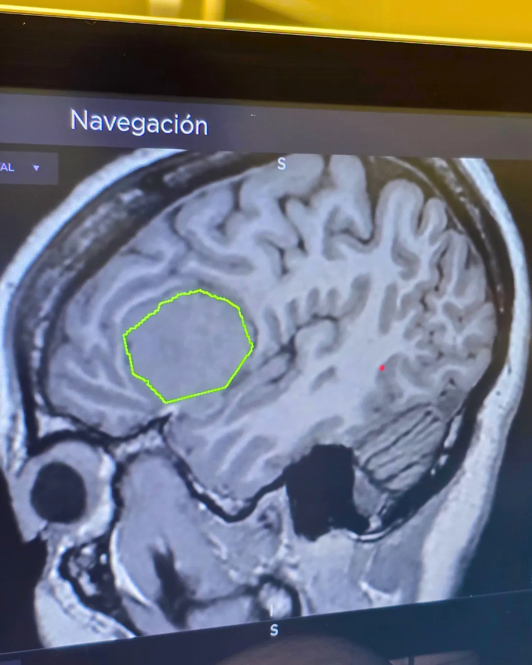
"Glioma" is not one diagnosis. That single word covers tumours with radically different outlooks.
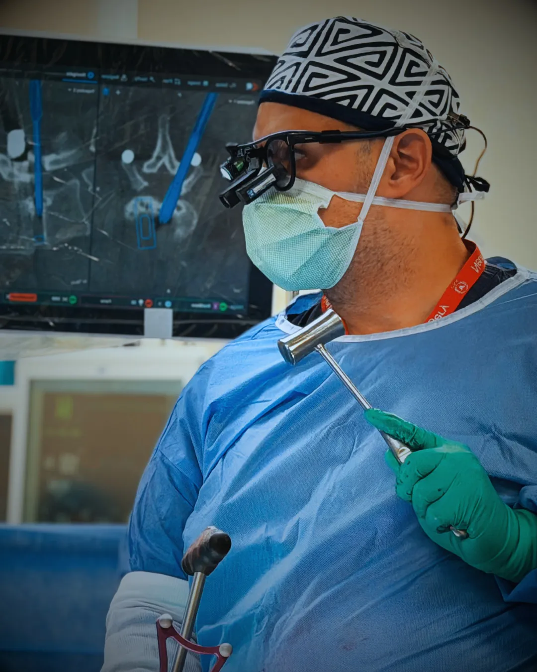
The most common brain tumour in adults — and the one whose outlook has changed most in the last decade.