
Lumbar disc herniation: when surgery is needed and when it isn't
The question is not whether you have a herniation. It is whether that herniation explains your symptoms, and whether time will resolve it.
It is not "laser surgery" and it is not risk-free. It is the same operation, performed through a smaller door.
Minimally invasive spine surgery — MIS — covers a set of techniques aiming to achieve exactly the same goal as conventional open surgery, but with far less trauma to the healthy tissue you have to cross to get there.
In classic open surgery, reaching the spine requires stripping and retracting the paraspinal musculature from the midline and holding it retracted throughout the operation. That muscle is precisely what stabilises the spine. The damage it sustains explains much of the postoperative pain and prolonged recovery.
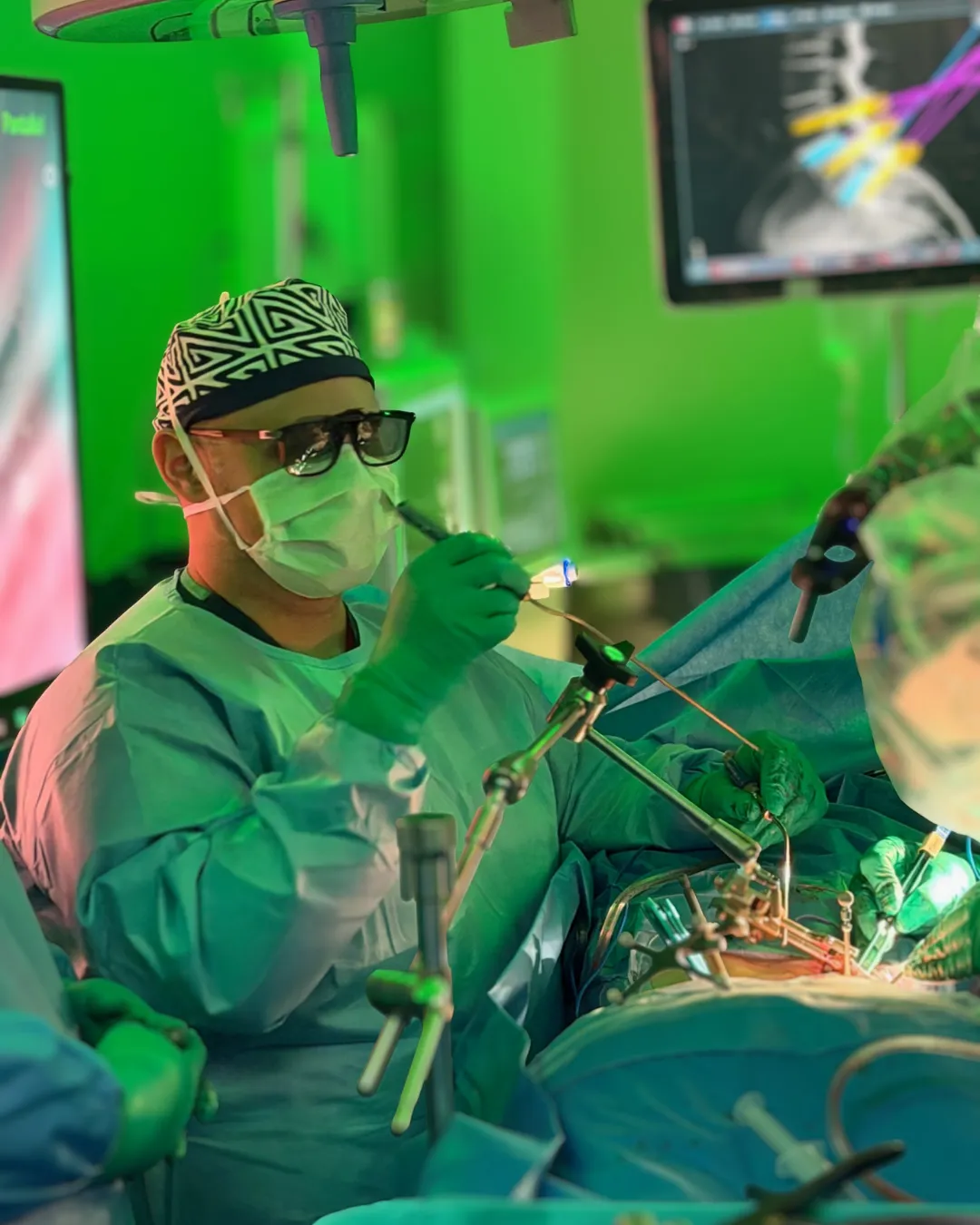
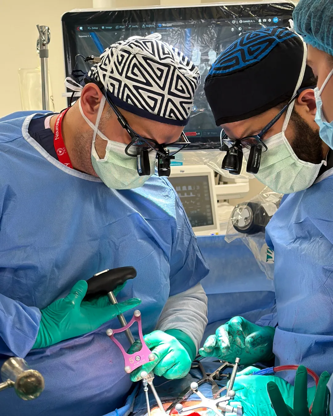
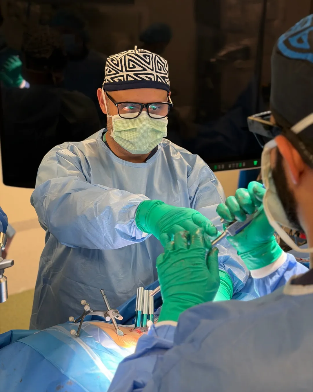
In minimally invasive technique a tubular retractor is used: muscle fibres are progressively dilated to create a working corridor, without cutting them or stripping them off the bone. Surgery is performed through that tube, with optical magnification and navigation.
| Open surgery | Minimally invasive | |
|---|---|---|
| Incision | Typically 8–15 cm | Typically 2–4 cm |
| Musculature | Stripped and retracted | Dilated between fibres |
| Blood loss | Greater | Less |
| Postoperative pain | Greater | Less |
| Hospital stay | Longer | Shorter |
| Surgeon's view | Wide, direct field | Narrow field, requires magnification and navigation |
| Learning curve | Standard | Considerably more demanding |
That last row matters more than it looks: MIS technique demands specific training and surgical volume. It is not simply "making the incision smaller".
For lumbar disc herniation. Through a tube a few centimetres wide, the fragment compressing the nerve root is removed. It is the most common MIS operation and the one with the fastest recovery.
For lumbar spinal stenosis. It widens the canal and frees the nerves, even bilaterally while working from one side only, preserving the structures on the opposite side.
Transforaminal interbody fusion by minimally invasive route. Access is from behind and through the foramen; the damaged disc is removed, a spacer restoring height is placed, and fixation is achieved with percutaneous screws. Indicated in spondylolisthesis and instability.
Lateral approach: the spine is reached from the patient's side, through the psoas muscle, without touching the posterior structures or the great anterior vessels. It allows placement of a large spacer that corrects height and alignment very effectively. It requires continuous neuromonitoring of the lumbar plexus during access.
The most extreme expression of the MIS philosophy: an endoscopic camera and instruments work through an incision of under a centimetre. Applicable to selected cases of disc herniation and foraminal stenosis.
Pedicle screws are placed through small stab incisions, guided by 3D navigation or fluoroscopy, without exposing the whole spine.
A surgeon who commands both techniques chooses the right one for each case. The correct answer is never "everything is done minimally invasively".
| Procedure | Usual stay | Desk work | Full activity |
|---|---|---|---|
| Microdiscectomy | 0 – 1 day | 2 – 4 weeks | 6 – 12 weeks |
| Tubular decompression | 1 – 2 days | 3 – 5 weeks | 8 – 12 weeks |
| MIS-TLIF / XLIF | 2 – 4 days | 4 – 8 weeks | 3 – 6 months |
These are indicative ranges: actual progress depends on age, baseline condition, the number of levels operated and associated medical conditions. An individual timeline is provided in consultation, not an average.
A good surgeon answers these in detail and without discomfort. Question two is the most revealing: if nobody explains what happens if you don't have surgery, half the conversation is missing.
It has clear advantages in blood loss, postoperative pain, hospital stay and wound infection. But it retains the risks inherent to operating near neural structures and demands a considerably steeper learning curve. Safety depends as much on the correct indication and the team's experience as on the technique itself.
No. "Laser spine surgery" is largely a commercial term; lasers have very limited and specific applications in spine surgery. Minimally invasive surgery uses tubular retractors, optical magnification and intraoperative navigation — not a laser that dissolves the herniation.
Most lumbar herniations are candidates for tubular microdiscectomy. The exceptions are herniations in difficult locations, revision cases with abundant scar from previous surgery, or anatomies where a tubular corridor does not allow safe access. The decision is made case by case.
Not in most microdiscectomies or simple decompressions. Screws are placed only when there is demonstrated instability, significant spondylolisthesis, deformity, or when the required decompression would compromise the stability of the operated segment.
Dr. Ricardo Bermúdez operates at Hospital Punta Pacífica (Pacífica Salud) and The Panama Clinic in Panama City, in operating rooms equipped with neuronavigation, intraoperative imaging and neuromonitoring. Consultations can be booked via WhatsApp at +507 6202-7644.
Written and reviewed by Dr. Ricardo Bermúdez, neurosurgeon. Last reviewed: 2026-09-09.
General informational content. It is not medical advice, does not create a doctor-patient relationship, and must not be used to self-diagnose or to decide on treatment. Outcomes vary from person to person. If you have symptoms, see a doctor.
The question is not whether you have a herniation. It is whether that herniation explains your symptoms, and whether time will resolve it.
A characteristic pattern: walking a distance, having to stop, and being able to continue after sitting for a moment.
A slipped vertebra sounds alarming in a report. In practice, many grade I cases never cause trouble.