

Lumbar spinal stenosis: when walking becomes the problem
A characteristic pattern: walking a distance, having to stop, and being able to continue after sitting for a moment.
A slipped vertebra sounds alarming in a report. In practice, many grade I cases never cause trouble.
Spondylolisthesis is the forward slippage of one vertebra over the one immediately below it. It almost always occurs in the lower lumbar spine: L4 over L5, or L5 over S1.
It is a finding that alarms people when it appears in a radiology report. Context helps: many people live with mild spondylolisthesis for decades without any symptoms.
The most common. It appears after age 50–60, more often in women and typically at L4-L5. The facet joints and disc wear out, lose their holding capacity, and the vertebra slips. It is almost always accompanied by lumbar spinal stenosis.
Caused by a defect in the pars interarticularis, the bone bridge connecting the facets. It usually originates in adolescence, often in athletes performing repeated hyperextension — gymnastics, diving, football — and can stay silent for years. Typical at L5-S1.
| Grade | Slippage | Behaviour usually described in the literature |
|---|---|---|
| I | 0 – 25% | The most common. Many are asymptomatic. Conservative management first. |
| II | 25 – 50% | Often symptomatic. May need surgery if nerves are compressed. |
| III | 50 – 75% | Usually symptomatic. Surgery clearly enters consideration. |
| IV | 75 – 100% | Significant deformity. Surgery in most cases. |
| V (spondyloptosis) | > 100% | The vertebra has dropped in front of the one below. Rare and complex. |
To repeat the important point: grade alone does not decide surgery. A grade I with severe stenosis and disabling pain is operated on sooner than an asymptomatic grade II found incidentally.
A clinically useful detail: patients often say they walk better pushing a shopping trolley or going uphill. Leaning forward opens the canal and eases symptoms.
Many patients stabilise here and need nothing more. Interestingly, in degenerative spondylolisthesis the slippage tends to self-stabilise over time as the segment stiffens.
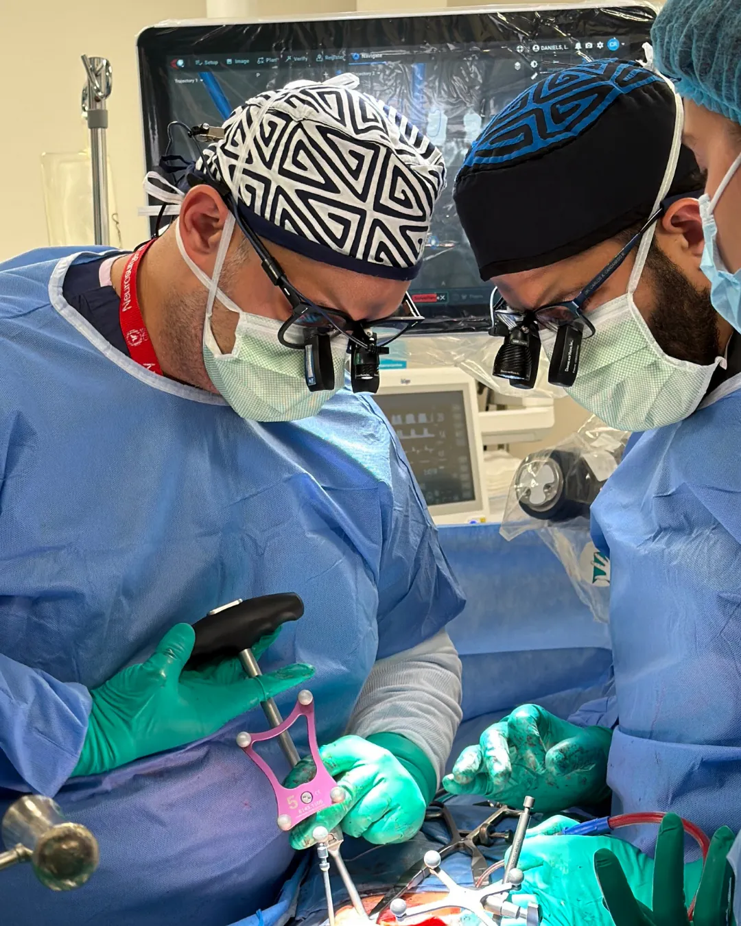
Today much of this surgery is performed with minimally invasive technique and 3D navigation: the position of each screw is verified with imaging inside the operating room before finishing, and muscle is split rather than cut. That means less blood loss and shorter recovery than classic open surgery.
Decompression relieves leg pain relatively quickly. Bone fusion, by contrast, is a biological process taking months: bone must grow and unite the vertebral bodies. The postoperative course therefore has phases:
Smoking is the modifiable factor that most affects fusion: it significantly reduces the chance that bone will consolidate. Stopping smoking before a fusion is not generic health advice — it is part of the treatment.
Not necessarily. In the degenerative form, slippage usually progresses little and often tends to self-stabilise as the segment stiffens. In the isthmic form in adolescents, imaging follow-up is advisable because it can progress during growth. Progression is monitored with serial X-rays.
Yes, with adaptations. Core and stabiliser strengthening is recommended, while avoiding repeated lumbar hyperextension and heavy axial loading. Walking, swimming, stationary cycling and motor control work are usually well tolerated. Ideally a physiotherapist should adapt the programme to your grade and symptoms.
Almost never as a first step. Most grade I cases are managed conservatively and many are asymptomatic, discovered incidentally on an X-ray ordered for another reason. Surgery is considered if there is symptomatic nerve compression, demonstrated instability, or disabling pain not responding to treatment.
Spondylolysis is the defect or stress fracture in the pars interarticularis, without the vertebra having slipped. Spondylolisthesis is the slippage itself. Spondylolysis can progress to spondylolisthesis if the defect allows the vertebra to slide, but this does not always happen.
It depends on the type of work. For office work, many patients return between 4 and 8 weeks. For physical work involving lifting, the timeframe usually extends to 3–6 months, conditional on fusion consolidating, which is verified with imaging. The schedule is individualised at each postoperative review.
Written and reviewed by Dr. Ricardo Bermúdez, neurosurgeon. Last reviewed: 2026-09-09.
General informational content. It is not medical advice, does not create a doctor-patient relationship, and must not be used to self-diagnose or to decide on treatment. Outcomes vary from person to person. If you have symptoms, see a doctor.
A characteristic pattern: walking a distance, having to stop, and being able to continue after sitting for a moment.
In most low back pain no specific structural cause is identified. And most of it still gets better.
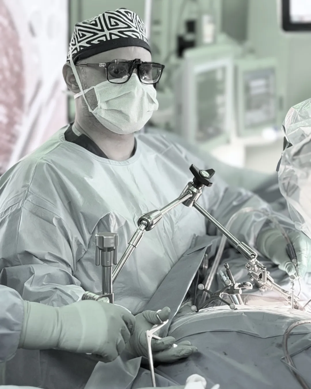
It is not "laser surgery" and it is not risk-free. It is the same operation, performed through a smaller door.